Test
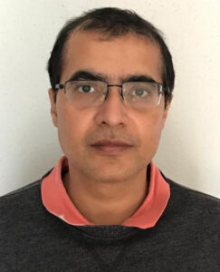
Aditya Mishra
Dr. Aditya Mishra is an environmental AI cluster hire faculty in the Department of Statistics. Prior to joining the department, Dr. Mishra was a computational/data scientist at the Platform for Innovative Microbiome and Translational Research at the University of Texas MD Anderson Cancer Center. At MD Anderson, Aditya was involved in developing and applying statistical methods to understand the role of the human microbiome in cancer onset, progression, and response to therapy. Aditya’s research mainly focuses on developing statistical methods and computational tools using the framework of High-dimensional statistics, Multivariate analysis, Reduced-rank regression, Regularization, Robust statistics, Multi-omics, Computational biology, Causal inference and Variational inference. He has extensive interdisciplinary research experience in a variety of fields, including microbiome data analysis, genomics, cancer genomics, public health, ocean microbiology, and dietary intervention study. As a part of environmental, Aditya will continue to understand and investigate the role of microbiome various context including cancer genomics, marine science, soil science and agriculture science.
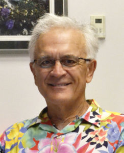
Alison Clune Berg
Alison's current research involves evaluating community Extension education programs to improve nutrition behavior for the prevention and management of chronic disease, including obesity, cardiovascular disease, diabetes, and some cancers.
Our research falls under the umbrella of exploring the effectiveness of the Extension model to enhance knowledge and facilitate healthy behavior change across the lifespan. Specifically, we focus on the impact of UGA Extension's Cooking for a Lifetime of Cancer Prevention on cancer preventive lifestyle behaviors and screening for breast, cervical, and colorectal cancer among urban and rural Georgians and exploring the relationship of program implementation factors on outcomes. Newer research includes exploring feasability and acceptability of the National Diabetes Prevention Program in UGA Extension and the impact on diet quality, physical activity, and physical function among middle-aged and older participants, and the relationship of health insurance status and preventative care behaviors on participant outcomes.
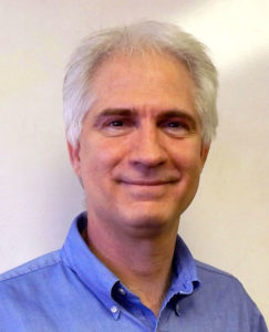
Bin Yi
Dr. Bin Yi is an Associate Research Scientist at the University of Georgia's College of Pharmacy, Department of Pharmaceutical and Biomedical Sciences. Currently, he serves as a Graduate Program Faculty member at the University of Georgia, contributing to the academic and professional development of students in the pharmaceutical sciences.
Dr. Yi’s research focuses on three main areas: 1. Developing novel therapeutic strategies for colon and breast cancer, both in vitro and in vivo. 2. Identifying and characterizing novel molecules in the tumor microenvironment that influence cancer progression. 3. Studying exosomal miRNAs and their role in cancer metastasis.
In addition to his research, Dr. Yi is dedicated to teaching and mentoring students at various academic levels, guiding them through research projects and scientific presentations. He also plays a vital role in departmental service, participating in numerous committees and review panels. Dr. Yi's commitment to advancing cancer research and education makes him a valuable asset to the scientific community.
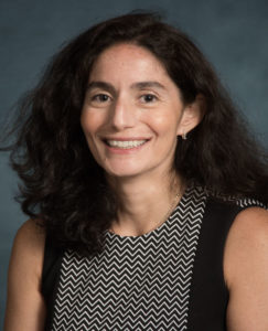
Corey Saba
Oncology Service, Small Animal Medicine & Surgery, Veterinary Teaching Hospital
Professor
EXPERTISE: Oncology
BIOGRAPHY:
Educational Background
2006 Diplomate American College of Veterinary Internal Medicine (Specialty of Oncology)
2002 Doctor of Veterinary Medicine, Louisiana State University, School of Veterinary Medicine, Baton Rouge, LA
1996 Bachelor of Arts, Biology, Rhodes College, Memphis, TN
Teaching Experience
2012—present Associate Professor of Oncology, Department of Small Animal Medicine & Surgery, College of Veterinary Medicine, University of Georgia
2008—2012 Assistant Professor of Oncology, Department of Small Animal Medicine & Surgery, College of Veterinary Medicine, University of Georgia
2006—2008 Instructor of Oncology, Department of Small Animal Medicine & Surgery, College of Veterinary Medicine, University of Georgia
David J. Garfinkel
The Garfinkel lab studies the mechanism and consequences of Ty1 retrotransposition in the budding yeast Saccharomyces. Ty1-5 elements are similar to retroviruses such as HIV and a variety of retroelements widely dispersed in nature. We recently discovered a novel self-encoded derivative of the Ty1 capsid protein that modulates retrotransposition in a dose-dependent manner by inhibiting virus-like particle (VLP) assembly and function. The Ty1 restriction factor is similar to mammalian restriction factors derived from domesticated retroelement capsid genes that now defend their hosts from infection by oncogenic retroviruses. Ty element biology is a major paradigm for understanding other retroelements. For example, almost 50% of the human genome is comprised of retroelement sequences, such as LINE, SINE and endogenous retroviruses because utilizing RNA as a replication template can give rise to massive amplifications if left unchecked. Chromosomal rearrangements and insertional events involving these elements have been implicated in human disease and cancer. Several of these events were initially characterized at the molecular level and continue to be studied with Ty in the powerful yeast model. Since the retrotransposon and retroviral replication cycles are closely related, the process of Ty retrotransposition can be compared and contrasted with retroviruses to help illuminate areas of research more difficult to approach in other organisms. Furthermore, recent work shows that the neuronal Arc gene is derived from a Ty3-like element and encodes a capsid protein that facilitates synaptic communication by transmission of Arc VLPs containing Arc mRNA and perhaps additional cellular transcripts. Arc is associated with learning and long-term memory in Drosophila and mammalian models.
Edward T. Kipreos
I have extensive experience with cell cycle regulation and signaling using mammalian tissue culture cells and C. elegans as experimental model systems. My background has provided a strong foundation in cell cycle regulation. As a graduate student with Dr. Jean Wang at UCSD, I studied the v-abl and c-abl tyrosine kinases and cell cycle regulation in mammalian tissue culture cells (a, b). As a postdoctoral fellow with Dr. Ed Hedgecock at Johns Hopkins University, I studied the regulation of cell proliferation in C. elegans. During my postdoctoral work, I co-discovered the cullin gene family when I cloned the cul-1 gene and showed that it was required for cell cycle exit in C. elegans (c). Cullins form the scaffold for cullin-RING ubiquitin ligase (CRL) complexes. A central focus of my laboratory is on the functions of CRL complexes, which we study in both C. elegans and mammalian tissue culture cells. CRLs are involved in diverse cellular pathways, and this has allowed us to make important discoveries in multiple areas of biology. We have discovered: how CRL4CDT2 complexes regulate DNA replication-licensing factors to ensure that genomic DNA is replicated only once per cell cycle; how CRL2FEM-1 controls sex determination by targeting the degradation of a Gli-family transcription factor; and how both CRL4 and CRL2 complexes regulate the cell cycle and cell motility by targeting the degradation of CDK-inhibitors (d). Current studies that focus on signaling include: the discovery that a CRL2 complex targets RSK1 and RSK2 for degradation in mammalian cells; the negative regulation of Notch signaling by the CRL2LRR-1 complex; and the discovery of a CRL-regulated pathway that links insulin signaling and the control of mitochondrial morphology. In an unrelated project, we created the first primary culture system for C. elegans germ cells and used it to identify a specific bacterial folate as an exogenous signal that stimulates germ stem cell proliferation. We show that the folate signaling occurs independently of the folate’s role as a vitamin in one-carbon metabolism, suggesting a new signaling pathway. This research has implications for cancer because the folate receptor is overexpressed in many cancer cells and promotes cancer proliferation. Our current research includes studies on non-canonical folate pathways in human cancer cells.
Eileen J. Kennedy
Our laboratory is focused on translational research relating to allosteric regulation of kinases. The long-term goal of the lab is to develop constrained peptide scaffolds that can be used to probe or inhibit cell signaling events that are mediated by various proteins including kinases. We have focused on the development of cell-permeable research tools that can be applied to perturb or disrupt protein-protein interactions (PPIs) that are involved in allosteric regulation or signaling within a cellular environment. These chemically constrained peptides target key protein-protein interactions that either regulate kinase activity or function. We have applied this strategy to disrupt PPIs that regulate various kinases including EGFR and LRRK2 and have also regulated PPIs that mediate microsignaling complexes within cells including AKAP complexes.
Eugene Douglass
The Douglass Lab combines the tools of organic chemistry, biochemistry and computational biology to overcome the urgent problem of multi-drug resistance (MDR) in cancer. Typically, MDR is caused by ~30 drug efflux and metabolism enzymes which can create 30 different types of “armor” for late stage cancers. Our Organic Chemistry program seeks to modify current chemotherapies to become “armor piercing”, our Biochemistry program seeks to characterize each “armor” substrate specificity (kcat/Km) and our Data-Science programs focuses on building clinical diagnostics to match drugs to individual patients.
Fikri Avci
Dr. Fikri Avci is currently Associate Professor of Biochemistry and Molecular Biology at the Center for Molecular Medicine in the University of Georgia. Dr. Avci is a glycoimmunologist who has training in both chemistry and biology of complex carbohydrates. He received his Ph.D. in 2005 from Rensselaer Polytechnic Institute. Subsequently, he worked as a postdoctoral research associate (2006-2011) and a faculty member (2011-2013) in the Departments of Medicine and Microbiology and Immunobiology at Harvard Medical School. He currently runs an interdisciplinary research group at the interface of carbohydrate research and immunology with an objective to explore treatment of and protection from infectious diseases and cancers by understanding key molecular and cellular interactions between the components of the immune system and carbohydrate antigens associated with microbes or cancers.
Gerald Hart
Georgia Research Alliance William Henry Terry, Sr. Eminent Scholar in Drug Discovery, and Professor of Biochemistry and Molecular Biology
We discovered O-GlcNAcylation (OGN) in the early 1980s. OGN is the cycling of N-acetylglucosamine on and off serine or threonine residues on nuclear and cytoplasmic proteins. OGN occurs on over four-thousand proteins where it serves as a nutrient sensor with extensive crosstalk with phosphorylation to regulate many cellular processes, including signaling, transcription, translation and mitochondrial functions. There is a rapidly growing literature suggesting that O-GlcNAcylation contributes to the properties and progression of cancer cells. O-GlcNAc cycling is universally elevated in cancer cells and, indeed, preventing increased O-GlcNAcylation can block cancer progression. Aberrant expression and activities of O-GlcNAc cycling enzymes, especially OGT, have been reported in all human cancers studied to date. Altered cellular metabolism is a major hallmark of cancer. Glucose uptake and glycolysis are accelerated in cancer cells (“Warburg Effect”), which gives cancer cells an advantage for intensive growth and proliferation. O-GlcNAc-dependent regulation of signaling pathways, transcription factors, enzymes, and epigenetic changes are all likely involved in metabolic reprograming of cancer. Several researchers have proposed that inhibition of hyper-O-GlcNAcylation could be a potential novel therapeutic target for cancer treatment. It has also been proposed that aberrant O-GlcNAcylated proteins might be novel biomarkers of cancer.
Currently, our laboratory is focused on the roles of OGN on -catenin in the functions of WNT signaling and in the epithelial-mesenchymal transition (EMT). In collaboration with Michael Pierce’s laboratory we are also investigating the roles of OGN in mechanisms of tumor cell suppression of the immune system.
Hang Yin
The research in Yin laboratory focuses on adult stem cell populations in skeletal muscle and adipose tissues. We use molecular genetic tools, and small molecule compounds to identify therapeutic targets/agents for skeletal muscle atrophy, injury, muscular dystrophies, obesity and type 2 diabetes. My laboratory uses various transgenic mouse models and primary cell cultures to understand gene functions and to model human diseases. We are particularly interested in characterizing functions of metabolism-related genes, epigenetic/chromatin factors, RNA-binding factors and non-coding RNAs in controlling adult stem cell behaviors (lineage determination, proliferation, differentiation, etc.).
Houijan Cai
I am interested in fatty acylation modification of proteins and its functional role in tumor progression. My early study demonstrated that protein palmitoylation is essential for the activity of Src family kinases. Gain of palmitoylation sites inhibits Src kinase activity, and loss of palmitoylation enhances Fyn kinase activity (Cai et al., PNAS, 2011). Since I joined UGA in 2013 as a tenure track faculty, my lab continuously focuses on how myristoylation and palmitoylation regulate Src family kinases, subsequently modulating their activity and oncogenic signaling. Thanks to the funding supports of the DOD and NIH, my lab investigated how dietary fatty acids regulates the activity of Src kinase and oncogenic potential. My lab has established experimental approaches of monitoring the biosynthesis of acyl-CoAs (Yang et al., Analytical Chemistry, 2017). We have clearly demonstrated that myristoylation is essential for Src mediated and high fat diet accelerated tumor progression. Myristoylation regulates the association of myristoylated Src in the cell membrane, its mediated kinase activity (Kim et al., JBC, 2017), and oncogenic signaling (Li et al., Neoplasia, 2018; Li et al., JBC, 2018). In addition, we have identified that a myristoyl-CoA analog, B13, and its derivatives could inhibit protein myristoylation and Src kinase activity (Kim et al., Cancer Research, 2017). Additionally, myristoylation is also essential for myristoylated proteins to be encapsulated into extracellular vesicles/exosomes. We extend our study to detect aggressive prostate tumors by monitoring myristoylated Src kinase in extracellular vesicles from blood plasma. We will potentially provide a non-invasive approach to detect the initiation of aggressive/metastatic tumors for the patients under active surveillance.
J. Michael Pierce
Dr. J. Michael Pierce received his Ph.D. from the Johns Hopkins University Department of Biology, and he completed an NIH Postdoctoral Fellowship at the Univ. of California, Berkeley, Dept. of Biochemistry with Dr. Clinton Ballou. He was appointed an Assistant Professor in the Department of Cell Biology, Univ. of Miami Medical School in 1982, and received the 5-year Faculty Research Award from the American Cancer Society in 1988. After moving to the Univ. of Georgia in 1991, he has received the Distinguished Research Professor award, and serves as the endowed Mudter Professor of Cancer Research. He is also the Director of the University of Georgia Cancer Center and is Principal Investigator of the National Center for Biomedical Glycomics, an NCI U01 award focusing on cancer glycomarkers, as well as the NIGMS T32 Glycoscience Training Program for Graduate students, the only training program of its kind in the U.S. He is the Chair of the board of the non-profit East Georgia Cancer Coalition, which focuses on delivering cancer prevention and screening to underserved populations in eastern Georgia. His research focuses on the application of monoclonal antibody technologies to find glycoprotein targets for cancer therapies.
Jeffrey L. Urbauer
Since 1992, my research has focused on the study of proteins and protein-protein interactions using molecular biology, biophysical methods, and, in particular, biomolecular NMR spectroscopy, as major research tools. My expertise is in protein chemistry and biomolecular NMR spectroscopy, having worked in the former for more than 30 years and the latter for over 20. I’ve played central roles in establishing high field biomolecular NMR facilities, including all aspects of instrumentation evaluation, purchase, installation and facility management, at the State University of New York at Buffalo (Chemistry) and the University of Pennsylvania (Biochemistry and Biophysics, School of Medicine), in both cases as a research (non-tenure track) professor, and in association with Professor A. Joshua Wand. As a first-year, tenure-track assistant professor at the University of Kansas (KU), I was PI for a successful proposal to the NSF that provided funding for the acquisition of a 600 MHz NMR system for biomolecular research, and was responsible for administering that project. Before leaving KU, I was heavily involved in efforts to establish a high field (800 MHz) NMR facility, including instrument evaluation, purchase and facility design. At the University of Georgia, I served as one of the focus group coordinators for the NIH-sponsored Southeast Collaboratory for High-Field Biomolecular NMR and also as the University Representative on the Governing Council for that facility. In 2010, I was the PI of a successful proposal to the NIH Shared Instrument Grant program for a new electronics console for our 800 MHz NMR instrument. I serve routinely as a reviewer grant proposals to NIH instrumentation programs, and as a reviewer for NIH and NSF grant proposals for structural biology, physical biochemistry and biomolecular NMR research.
Jia-Sheng Wang
My research interest is in studying etiology and prevention of environmental toxicants and pathogens, especially mycotoxins, and related human diseases with major focus on cancer and other chronic diseases. On the national scale my research involves molecular biomarkers, molecular epidemiology, cancer chemoprevention, and toxicology, and internationally I have conducted research in various areas including in countries Burkina Faso, China, Ghana, Indonesia, Malaysia, Mexico, and Uganda for etiology and prevention of cancers and other chronic diseases. I have conducted human clinical and chemoprevention trials for almost 25 years. I have developed strong research program in genomics, metabolomics, and microbiome research over past 15 years.
Jin Xie
The Xie Lab is interested in nanoparticle technologies. We engineer electrolyte-, polymer-, protein- and cell-based nanoplatforms that aim to solve unmet clinical needs. In particular, we are focused on developing novel nanotechnologies for cancer drug delivery, radiotherapy, imaging, and immunotherapy. Projects include salt nanoparticle therapeutics, X-ray induced photodynamic therapy, cell-based drug delivery, magnetic nanoparticle contrast agents, nanoparticle-based immunotherapy, and nanoparticle radiosensitizers.
Jon Mochel
Dr. Jon P. Mochel obtained his Veterinary Medical Degree from the National Veterinary School of Alfort (Paris, France). He completed his Doctorate studying Neurosciences in collaboration with the College de France and received the Silver Medal from Paris XII for his work. Dr. Mochel holds a MS in Pharmacology and Pharmacokinetics and is a Diplomate of the European College of Veterinary Pharmacology and Toxicology (ECVPT). He completed his Ph.D at Leiden University (Netherlands), with a focus on the mathematical modeling of the cardiovascular system for optimization of ACE inhibitors dosing schedules in patients with congestive heart failure. He is a founder of the Animal Health Modeling & Simulation Society which aims at promoting the dissemination of mathematical modeling in Veterinary Sciences. Dr. Mochel is a Professor of Systems Pharmacology in the Department of Pathology at the University of Georgia and a Fellow of the American Academy of Veterinary Pharmacology and Therapeutics. He is an ad hoc Study Section Reviewer for the US National Science Foundation, the UK Medical Research Council and the CNRS/INSERM ATIP-Avenir Program. He is currently the President of the European Association of Veterinary Pharmacology and Toxicology and the recipient of the 2022 Research Award from the American Academy of Veterinary Pharmacology and Therapeutics. His research pertains to the analysis of (pre)clinical data obtained from spontaneous animal models of human diseases to bridge the knowledge gap between experimental models and patients. He has authored more than 130 peer-reviewed publications in select biomedical journals and is the co-founder of LifEngine Animal Health, a Y Combinator company developing gene editing solutions for canine oncology, and 3D Health Solutions, a platform for efficient drug screening in pharmaceutical research.
Karin Allenspach Jorn
Karin Allenspach received her veterinary degree from the University of Zurich. She did an internship in small animal emergency medicine and critical care at Tufts University and a residency in small animal internal medicine at the University of Pennsylvania and is a Diplomate of the European College of Veterinary Internal Medicine. She was awarded a PhD in veterinary immunology from the University of Bern, Switzerland for her work on canine chronic enteropathies. She is currently employed as Professor of Comparative Medicine at the University of Georgia, Athens, GA, and is a PI of the SMART Translational Medicine Lab at UGA, which focuses on the development and culture of adult stem stem-cell-derived organoids from various species. Karin is widely published and has a current H factor of 43. She has been internationally involved in several large-scale efforts to change governmental policies for veterinary medicine, for example by being a member of the ENOVAT group which is providing language for antibiotic use in dogs with diarrhea for all European countries. Furthermore, she has been a Board member of both the European and Comparative Gastroenterology Societies, as well as being president of the European Congress of Veterinary Internal Medicine for 12 years. Her latest efforts have resulted in the founding of a start-up company (3D Health Solutions, Inc.) with the goal of commercializing assays for drug screening based on organoid methods.
Kevin K. Dobbin
Biostatsisticians are critical for translating new cancer discoveries into clinical tests and assays that can benefit patients. Dr. Dobbin’s focus is the are of treatment selection biomarkers, which are biological measurements used to make clinical decisions about which treatment a patient should receive. His research also spans into multiplex and high dimensional assays, and related issues of reproducibility and computational methods.
Kosuke Funato
My research focuses on glioblastoma, the most aggressive brain tumor and one of the most lethal tumor types in both adults and children. I received my Ph.D. for my research on glioblastoma stem-like cells and have been involved in glioblastoma research for more than 10
years.
In the recent years, I have been studying recurrent histone mutations found in pediatric glioblastoma: histone H3K27M and G34R/V mutations. It has been shown that these two types of histone mutations define clinically and molecularly distinct subtypes of glioblastoma.
However, little was known about their cell types of origin and the precise mechanisms by which the histone mutations transform particular cell types. To address these questions, I developed a new tumor model system based on human embryonic stem cells (hESC) (Funato et al., Science 2014; Funato et al., Cell Stem Cell 2021). Given that hESCs can be differentiated into various neuronal cell types by chemically-defined protocols, this system enables us to interrogate the role of each single mutation, or combination of mutations, in defined cell populations. Our
studies demonstrate that the hESC-based tumor model is a powerful system to interrogate the genetic, molecular, and cellular mechanisms underlying gliomagenesis. In combination with other methodologies, such as patient-derived cell lines and genetic mouse models, our lab aims
to identify the subtype-specific mechanisms of gliomagenesis and develop tailor-made therapies for patients suffering from this devastating disease.
Lance Wells
My laboratory is focused on understanding the role that post-translational modifications, specifically glycosylation, play in increasing functional diversity of proteins. For the last 16 years, my lab has primarily focused on the biomedical significance of O-glycosylation, specifically the nutrient-sensing O-GlcNAc modification of nuclear and cytosolic proteins and the O-mannosylation pathway that is defective in many cases of congenital muscular dystrophy. The laboratory also has extensive experience in analytical mass spectrometry and the development/application of glycomic and glycoproteomic capture, identification, and quantification approaches. I serve as a Co-Director of the ThermoFisher Scientific appointed center of excellence in glycoproteomics that allows my laboratory to interact at both the software and hardware level with a leading instrument manufacturer to drive technology developments like those outlined in this proposal.
Leidong Mao
Leidong Mao, an associate professor in the College of Engineering, creates opportunities for students to conduct cutting-edge nanotechnology research. His current research interests include nanoscale magnetism and its application in biomedical applications such as cell sorting, single cell study, droplet microfluidics, hyperthermia, and magnetic nanomanipulations. He received the NSF CAREER Award in 2012, and a Young Scientist Award from 13th International Conference of Magnetic Fluids in 2013. Read more about Dr. Mao's teaching and research in Focus on Faculty.
Lohitash Karumbaiah
Dr. Karumbaiah joined the University of Georgia’s Regenerative Bioscience Center and Animal Science Department in October 2013, where he is currently associate professor of regenerative medicine. His research is focused on the design and development of novel interventional therapies to combat traumatic brain injuries and invasive brain tumors. Dr. Karumbaiah received his PhD in 2007 from the University of Georgia. He subsequently joined Prof. Ravi Bellamkonda’s group in the Wallace H. Coulter Department of Biomedical Engineering at Georgia Institute of Technology and Emory University School of Medicine, where he designed and developed novel biomaterials for neural tissue repair. His research interests include (1) gaining a better understanding the brain tumor microenviroment in order to develop targeted therapies to stem tumor invasion and enhance the efficacy of standard-of-care therapeutics; and (2) the development of novel glycomaterial scaffolds and functional electrical stimulation tools to help trigger the endogenous regenerative cascade and facilitate functional recovery following traumatic insults to the nervous system. His laboratory has developed novel glycomaterials, microfluidic devices, and functional electrical stimulation tools to answer fundamental questions related to these areas of study. Dr. Karumbaiah has previously worked in product development and on new product launch projects at Monsanto Company, and served as study director for a medical device company. His research is funded by multi-year grants from the National Institutes of Health (NIH), National Science Foundation (NSF) Engineering Research Center for Cell Manufacturing Therapies (CMaT), the Alliance for Regenerative Rehabilitation Research and Training (AR3T), and seed-grants from the Regenerative Engineering and Medicine (REM) partnership.
Bibliography: http://www.ncbi.nlm.nih.gov/sites/myncbi/lohitash.karumbaiah.1/bibliography/40232329/public/?sort=date&direction=ascending
Mary Goll
My research program uses zebrafish to answer questions about vertebrate chromatin that would be difficult to address in other animal or cell culture systems. Our primary focus is on a repressive form of chromatin called heterochromatin. I have 13 years of experience in zebrafish research. My group has published papers that use zebrafish at developmental stages from the early blastula to adulthood. I have demonstrated experience manipulating gene activity and protein function in zebrafish embryos through injecting mRNA, chemical inhibitors, and morpholinos. I also have expertise performing Tol2 mediated transgenesis and CRISPR/Cas9 mediated gene editing in zebrafish. In total, my group has already established 18 zebrafish lines with mutations in chromatin related genes. I have 20 years of experience working in chromatin biology. I have expertise with molecular approaches such as DNA methylation and chromatin immunoprecipitation assays. My group regularly combines these molecular approaches with cell-based approaches including immunohistochemistry and transmission electron microscopy. I have also developed fluorescent transgene reporters that enable in vivo monitoring of repeat induced transcriptional silencing across zebrafish development. My research program strives to utilize the unique combination of tools and skills available in our laboratory to open up underexplored research areas and to pursue the most impactful research questions possible. The creativity and adaptability of our research program is evident in my group’s publication record, which documents insights gained from distinct research projects all designed to maximize the advantages of zebrafish for the study of chromatin
Michael Tiemeyer
My laboratory has invested over 15 years in developing tools to perform structural analysis of major and minor glycans isolated from small amounts of material. We have also recently been focused on high-throughput, semi-automated annotation of mass spectral data, an essential tool for expanding the scope of glycomic analysis to biomedical and basic research applications. With our developed techniques we have made major contributions to glycomics and glycoproteomics of normal and diseased tissues. We have also established the Drosophila melanogaster embryo as a premier model system for functional glycomics. We now possess a deeper understanding of the full complement of N-linked, O-linked, and glycosphingolipid glycans expressed in the Drosophila embryo than for any other organism. Our methods for structural analysis are mature and completely translatable to investigating the glycomes and glycoproteomes of any tissue or cell type. In addition, a major component of my independent career has focused on functional analysis of glycans in various organisms and cellular systems (Drosophila, mouse, pathogens, human leukocytes). I have successfully utilized forward and reverse genetics, molecular cloning, and cell-based screening approaches to identify and characterize glycan binding activities, glycan-based developmental phenotypes, and glycan processing mechanisms. These experiences, as well as the full range of my training in carbohydrate structure/function provide me with the necessary expertise to contribute meaningfully to the goals of this proposal.
Nancy Manley
My lab is primarily focused on studying the "life history" of the thymus, the primary lymphoid organ responsible for the generation of T cells. This approach encompasses the evolution, fetal development, postnatal function, and aging of this critical organ. Our basic hypothesis is that these diverse aspects of the biology of the organ are controlled by common regulatory networks, cellular dynamics, and physiological processes. We also study the parathyroid, which is required for calcium homeostasis, and has a shared developmental ontogeny with the thymus. We use a variety of approaches to accomplish these goals, including genetic analysis of tissue-specific and inducible mutant mouse strains, comparative and experimental embryology, and immunological techniques. Several of our current projects include natural and induced cellular fate transformations to generate thymic epithelial cells from heterologous cell types, that are either natural sources of ectopic T cell generation, or may be sources of thymus organs for transplant.
Natarajan Kannan
My research program is focused on deciphering the evolutionary rules governing the structure, function and regulation of complex cellular signaling systems, in particular protein kinases, using a combination of computational and experimental approaches. Important goals are to identify genomic variants associated with protein kinase regulation in disease and normal states and to develop computational resources for integrative mining of protein kinase data.
Unique qualifications: I am one among the very few investigators using a combination of computational and experimental approaches to investigate the evolution of regulation in the protein kinase fold-family of enzymes.
I have made impactful contributions describing the unique sequence and structural features of various protein kinase groups and families. My papers are well cited (over 2500 citations) and have provided a framework for understanding the complex modes of protein kinase regulation in diseases such as cancer. In recognition of my work, I have been invited to speak at major international meetings, serve on review panels, and co-organize international meetings on kinases. We have recently begun to study the evolution of glycosyltransferases and proteases using the specialized tools and resources developed for the study of protein kinases. I believe that that common evolutionary themes will emerge by studying these large enzyme super-families in parallel.
Collaborative team: As evidenced through my publications, I have a long and successful history of collaborative research with both domestic and international scholars. Collaborators include protein kinase biochemists, cell biologists and structural biologists. We have also collaborated with large-bioinformatics consortiums to develop new resources and annotations for the signaling community and are beginning to work with the glycobiology community to develop specialization tools for the study of glycoenzymes.
Robert S. Haltiwanger
My laboratory identifies and characterizes O-linked glycans in a wide variety of contexts. My initial work concentrated on O-GlcNAc, in which I identified proteins bearing the modification, mapped sites of modification, and identified the enzyme that adds O-GlcNAc to proteins (OGT). In my own laboratory, I have focused on O-fucose and O-glucose glycans on Epidermal Growth Factor-like repeats (EGF repeats) and Thrombospondin Type 1 Repeats (TSRs). This research has contributed greatly to the identification and characterization of the unique enzymes required for synthesis of these glycans, including demonstrating that the Fringe family of Notch modulators are 3-N-acetylglucosaminyltransferases, molecular cloning, expression and characterization of POFUT1 and POFUT2, initial identification and characterization of the 3-glucosyltransferase modifying O-fucose on TSRs, demonstrating that the Notch-pathway gene Rumi encodes POGLUT1, and identifying the two xylosyltransferases responsible for elongating O-glucose. We recently identified two novel enzymes that add O-glucose to a novel site on EGF repeats: POGLUT2 and POGLUT2. We have all of these enzymes overexpressed and have used them to glycosylate EGF repeats and TSRs in vitro in sufficient quantities for structural studies (NMR and/or X-ray crystallography). In collaboration with others, we have recently solved the structure of three of these enzymes co-crystallized with their acceptor substrates: Rumi/POGLUT1 with an EGF repeat, XXYLT1 with and EGF-O-Glc-Xyl, and POFUT2 with a TSR. We have developed glycomic methods for analyzing O-fucose and O-glucose structures and glycoproteomic methods for site-specific O-glycan analysis on EGF repeats and TSRs. We mapped sites of glycosylation on mouse Notch1 and demonstrated which sites are necessary for Fringe enzymes to mediate their effects. We have also begun to use what we have learned about Notch glycosylation to develop inhibitors of Notch activity. In particular, we have demonstrated that fucose analogs can be used to inhibit Notch activity in a ligand-specific manner. This is the first example of a small-molecule inhibitor that functions in a ligand-specific manner.
Ryan Weiss
Dr. Ryan Weiss received his B.S. in chemistry in 2008 at Point Loma Nazarene University in San Diego, CA. He then received his Ph.D. in chemistry in 2015 at the University of California, San Diego under the supervision of Prof. Yitzhak Tor. He then moved to the Department of Cellular and Molecular Medicine at the University of California, San Diego, where he worked as a postdoctoral fellow in the laboratory of Prof. Jeffrey Esko. Dr. Weiss began his independent career as an assistant professor in the Department of Biochemistry and Molecular Biology at the University of Georgia in January 2021. His current research interests include drug discovery and using genomic tools to understand the regulation of glycosylation in human diseases.
Shaying Zhao
I have been focusing on dog-human comparative genomics and oncology research since 2005. I am the first person to propose and use a novel dog-human comparison strategy for cancer driver-passenger discrimination, a central aim of cancer research. Funded by the National Cancer Institute, the American Cancer Society and the Georgia Cancer Coalition, work from my lab has shown a strong dog-human molecular homology for histologically matched cancer types/subtypes for mammary cancer, colorectal cancer, and head and neck squamous cell carcinoma. My lab has successfully used this novel dog-human comparison strategy for cancer driver-passenger discrimination for amplified or deleted genes in human colorectal cancer (Oncogene 33:814-22). My lab has published the first comprehensive characterization of canine mammary cancers with whole genome sequencing (WGS), whole exome sequencing (WES), and RNA-seq analysis (Cancer Res. 74:5045-56). The same is true for canine head & neck cancer (PLOS Genetics, 11(6):e1005277), colorectal cancer and extreme intestinal polyposis that resembles human familial adenomatous polyposis (FAP). Funded by the American Kennel Club Canine Health Foundation and the English Cocker Spaniel Club of Northern California, my lab is searching for germline mutations that predispose dogs to cancer development, and to improve canine gene annotation by RNA-seq.
I was trained in biochemistry in the laboratories of Dr. Stephen Ragsdale and Dr. Aziz Sancar, a Nobel laureate. I have worked at The Institute for Genomic Research (TIGR) from 1998-2005, where I led the best bacterial artificial chromosome (BAC)-end sequencing operation in the world and made notable contributions to the sequencing of the human, mouse, rat, and cow genomes. In addition, I have built a comprehensive BAC resource database and a BAC end sequence search Web server (Nucleic acids research 28, 129-132). My lab currently consists of 6 PhD students, 4 in bioinformatics and 2 in biochemistry. We have built state-of-the-art data analysis pipelines and published a software tool for copy number analysis with WGS data. I have served on various NIH Cancer Study Sections. I served on an Expert Panel to evaluate the NCI Informatics Technology for Cancer Research (ITCR) program in 2018. I am currently serving on the Steering Committee of the NCI Integrated Canine Data Commons (ICDC) database.
Somanath Shenoy
I am a biomedical scientist specialized in translational research in urological cancers, prostate cancer (PCa) metastasis, tumor microenvironment and other lung diseases. My current research includes mechanisms regulating cellular plasticity in epithelial-to-mesenchymal transition (EMT), endothelial-barrier regulation and Endothelial-to-mesenchymal transition (EndMT) in PCa metastasis. The foundation for my research is built through several Intramural grants, Veteran’s affairs, American Heart, American Legion, NIH, and Department of defense, as Principal- or co-investigator. While my forte is laboratory research in experimental therapeutics, one of my major accomplishments has been the creation of a broad, multi-disciplinary research training program encompassing postdoctoral fellows, graduate and professional students, and residents that extend from the laboratory to the bedside. This training environment is significant as it educates people to be successful in the changing landscape of biomedical research. My trainees are able to move into their respected health care fields as practitioners and researchers with a firm mutual understanding of each other’s knowledge. I am also a NIH KL2 and TL1 core co-director representing UGA in the Research Education Executive Committee in its alliance with Emory, Georgia Tech, and Morehouse SOM that form the Georgia Clinical and Translational Science Alliance (Georgia-CTSA). This VA Merit review grant proposal seeks to identify and characterize the role of selected microRNAs in prostate cancer EMT and metastasis.
Tamas Nagy
Research in the Nagy laboratory focuses on the role of small G proteins in mammary gland development and carcinogenesis. Various genetically engineered mouse lines are used as models in these investigations.
Timothy A. Bolger
Associate Professor in BMB who recently transferred to UGA from the Dept. of Molecular and Cellular Biology at the University of Arizona. My lab’s research focuses on RNA biology, specifically the regulation of RNA-protein interactions during protein translation. In addition to the importance of translation regulation in cancer generally, one project in my lab also has a direct cancer focus, examining the impact of mutations identified in an RNA helicase in the brain cancer medulloblastoma. While at UArizona, I was a member of the NCI-designated Arizona Cancer Center and a trainer in the Cancer Biology graduate program there. I also previously held an American Cancer Society Research Scholar Grant and served on an ACS peer review panel for several years. I found these involvements to be helpful in advancing our research by interacting with other cancer researchers in various ways.
Vladimir Popik
Research Interests:
Photoswitchable bioactive compounds: Photoactivation of drugs, enzymes, and other bio-molecules allows for achieving of the spatial and temporal control of their action. We design and synthesize compounds that are inactive in the dark but are converted into bioactive form upon irradiation with light of an appropriate wavelength. One of our goals is to overcome the most common drawback of caged biomolecules, which are known to release bioactive substrates in the dark due to conventional or enzymatic hydrolysis. The non-photochemical reactions of photoactivatable compounds (hydrolysis, thermal decomposition, etc.) that we develop are very slow and do not result in activation of the substrate.
Two-photon photochemistry: UV and visible light is efficiently absorbed by muscle tissues and only red and near infrared (NIR) radiation has sufficient depth of penetration in our bodies. Photons in this region, however, have relatively low energy, limiting the range of processes they can initiate. We are working on the development of the precursors that can be converted into an active form using nonresonant two-photon excitation. This approach allows us to use red/NIR light for photoactivation of UV-chromophores, while avoiding substrates sensitive to visible light, and therefore reducing accompanying phototoxicity.
Photolabile protecting groups, known as “cages” in biochemistry, allow for the spatial and temporal control of substrate release, as well as “reagentless” deprotection. Our group designs novel types of "safety-catch" photo-cages suitable for protection of broad range of organic molecules.
Mechanisms of light-induced reactions are investigated using state-of-the-art time-resolved IR (TIR) and UV-Vis (TUV) spectrometers. This data is then used in the design of novel photoswitchable bioactive compounds.
Photo-"click" reactions: Connection (or ligation) of two fragments to make a larger molecule or structure, as well as labeling DNA, proteins, and even whole cells, are usually achieved with the help of the so-called “click chemistry”. This term describe a set of bimolecular reactions that meet the following criteria: reactions should be wide in scope but selective, produce high yield of the product, proceed with reasonable rate under mild conditions, and tolerate broad range of solvents. We work on the development of light-induced ligation techniques, which proceeds under ambient conditions and does not require additional reagents or catalyst.
Walter K. Schmidt
Ras oncoprotein activity is strongly influenced by post-translational modifications that occur at the COOH-terminus: isoprenylation, palmitoylation, proteolysis, and carboxylmethylation. Our studies are focused on the enzymes that direct these modifications and range from investigations of enzyme specificity to inhibitor development.
Biosketch: https://www.ncbi.nlm.nih.gov/myncbi/161WiFB1JY/cv/270848/
Bibliography: https://www.ncbi.nlm.nih.gov/myncbi/browse/collection/41088668/?sort=date&direction=descending
Wentao Li
The research in my laboratory combines biochemistry, genetics, adductomics and computational approaches to investigate effects of epigenetics and spatial genome organization on environmental carcinogen-induced DNA damage formation and repair. Specifically, we use next generation sequencing-based methods to identify the precise locations of DNA damage caused by environmental carcinogens (e.g., UV, cigarette smoke, and mycotoxin) and to measure the repair efficiencies at these locations on a genome-wide scale. We aim to identify the key factors that influence both the induction of DNA damage and the efficiency of damage removal by nucleotide excision repair. Understanding these mechanisms may improve the prevention strategies for environmental carcinogenesis and lead to the development of new tools for diagnosing and treating human cancers.
Yang Liu
Single Cell & Exosome Analysis
Dr. Liu’s research is centered around harnessing the capabilities of micro / nano-fluidic technologies to advance patient health outcomes. A pivotal aspect of his research program revolves around the development of microfluidic platforms with the purpose of revealing the intricate relationships between cellular phenotypes and molecular expressions at the single-cell level. This pursuit holds the potential to unveil novel insights into the fundamental mechanisms underlying cellular behavior and response.
Another significant focus within Dr. Liu’s laboratory is the integration of droplet microfluidics and protein electrophoresis techniques. This integration is geared towards the comprehensive analysis of proteoforms within individual exosomes. This intricate exploration offers the opportunity to unravel the complex landscape of protein variations and modifications, thereby contributing to a deeper understanding of disease pathways and potential therapeutic interventions.
Driven by a commitment to pushing the boundaries of knowledge, Dr. Liu’s research endeavors exemplify the cutting-edge applications of microfluidic technologies in unraveling the mysteries of cellular and molecular biology.
AREAS OF EXPERTISE
• Microfluidics
• Single cell / exosome analysis
• Micro / nano technology
Ying Xu
We are interested in elucidating non-genetic drivers of cancer formation, progression and metastasis through computationally mining and modeling omic data of cancer tissues. We are particularly interested in understanding how pH related stress drives metabolic reprogramming and how new metabolic exits created for some of the reprogrammed metabolisms may causally link to various phenotypic behaviors of a cancer, such as cell proliferation, drug resistance and cell migration.
Zachary A. Lewis
I established my laboratory at the University of Georgia (UGA) in 2011. We work with a variety of fungal species to achieve two major goals: (1) to understand assembly and function of specialized chromatin domains in the nucleus and (2) to develop new strategies to treat fungal infections. My laboratory at UGA is currently by the NIH, and we have received prior support from the American Cancer Society, the Department of Energy, and the March of Dimes Research Foundation. Our research has led to several important discoveries regarding the regulation and function of repressive chromatin domains, as well as other aspects of chromatin structure and function. Work in my own is focused primarily on the model eukaryote, Neurospora crassa, but we have also carried out genomics, epigenomics, and bioinformatics analyses of the medically relevant fungus Aspergillus nidulans, zebrafish, and a number of bacterial species through collaborations with others. Members of my lab are experienced using genomics, bioinformatics, genetics, molecular biology, and proteomics. We have also worked closely with the Meagher lab to develop and test targeted antifungal liposomes against several fungal pathogens. In addition to my talented and capable research team, I work closely with members of the UGA Fungal Biology Group (http://research.franklin.uga.edu/fungi/), which consists of over 15 research teams focused on many aspects of fungal biology. Our research team, environment, and ideal choice of experimental systems all facilitate rapid progress toward our research goals. In summary, my expertise in fungal biology will complement expertise in the adjoining Meagher laboratory and the Lin laboratory to ensure that the goals of this project are accomplished successfully and efficiently. I look forward to continuing as an active participant in our collaborative work to develop Dectin-targeted antifungal liposomes and Dectin-based one-step fungal diagnostics.
Zachary A. Wood
Glucuronidation is a major detoxification pathway in the Phase II metabolism of drugs, and is often the source of unfavorable pharmacokinetics or pharmacodynamics during clinical trials; the rapid clearance of a drug reduces bioavailability, and the resulting glucuronides can be toxic. In addition, some cancers have been shown to co-opt glucuronidation as an effective drug resistance mechanism. One strategy for regulating glucuronidation is controlling the availability of the essential substrate, UDP-glucuronic acid (UDP-GlcA). By reducing UDP-GlcA levels, it should possible to sensitize those drug resistant cancers that utilize glucuronidation.
Human UDP-glucose 6-dehydrogenase (hUGDH) catalyzes the NAD+ dependent oxidation of UDP-glucose to UDP-GlcA. In addition to its role in drug metabolism, UDP-GlcA is also the substrate for the biosynthesis of UDP-xylose, which acts as an allosteric feedback inhibitor for hUGDH. In addition to allostery, hUGDH undergoes substrate-induced hysteresis (a slow isomerization of the enzyme from an inactive state to an active conformation). My lab uses structural biology and enzymology to understand the allosteric and hysteretic mechanisms that control hUGDH activity. Our ultimate goal is to leverage these mechanisms to control UDP-GlcA levels.
